Background & Challenges
In life sciences and biomedical research, scientists face the challenge of imaging deep into living tissues while maintaining high resolution and minimizing photodamage. Conventional fluorescence microscopes often struggle with limited penetration depth, photobleaching, and insufficient signal-to-noise ratios, which hinder advanced studies in neuroscience, immunology, and drug development.
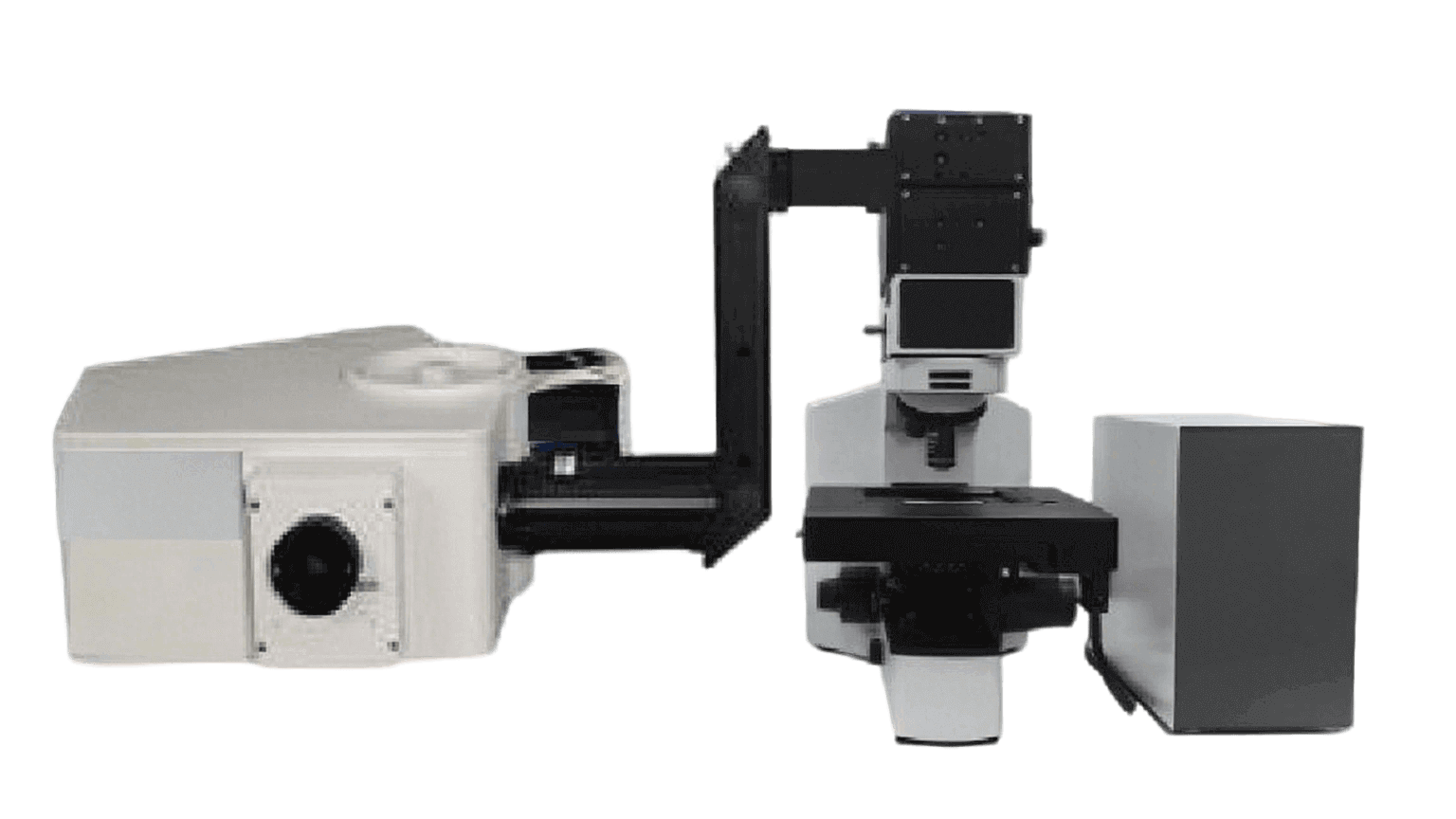
Solution: Multiphoton Microscope
The Multiphoton Microscope is an advanced fluorescence imaging system that leverages nonlinear optical processes, such as two-photon or three-photon excitation, to achieve high-resolution imaging deep inside biological tissues with minimal photodamage.
- Nonlinear Excitation: Two- or three-photon absorption enables efficient excitation at greater depths.
- High-Resolution Imaging: Scanning resolution ranges from 512 × 512 up to 4096 × 4096 pixels, supporting both macro- and micro-scale investigations.
- High-Sensitivity Detection: Equipped with a GaAsP PMT (Gallium Arsenide Phosphide Photomultiplier Tube), achieving quantum efficiency (QE) > 45%.
- Flexible Imaging: Variable zoom from 1× to 16× and imaging speed of 4 fps, enabling both detailed imaging and dynamic process observation.
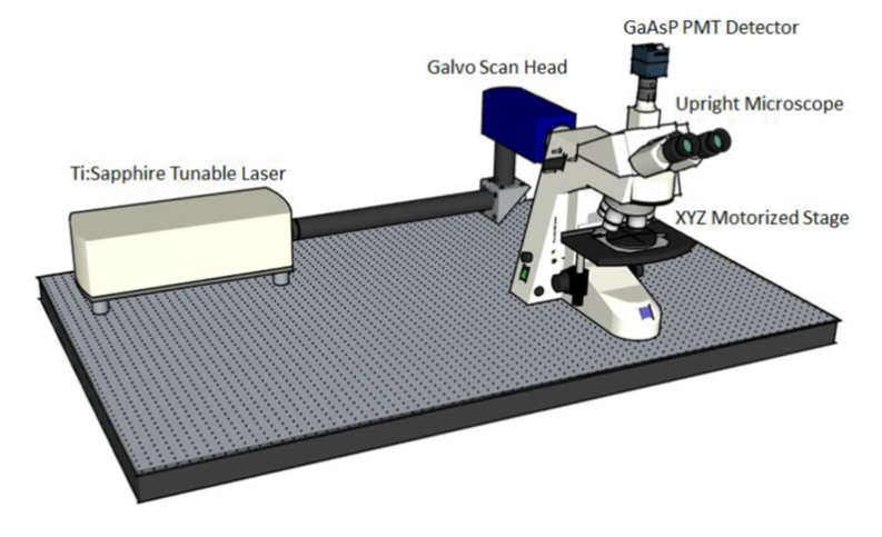
Customized Two-Photon System
To meet diverse research needs, the system supports flexible customization:
- Tunable Ti:Sapphire Laser: Covers wavelengths above 690 nm, with adjustable output power and pulse width of 100–150 fs.
- Core Scanning Module: Custom-designed optical housing and scanning module, ensuring system stability and adaptability.
- Microscope Platform: Includes widefield illumination system, eyepieces, objectives, condenser, and a motorized 3-axis stage for precise sample control.
Key Advantages
- Deep Tissue Imaging: Enables non-invasive imaging at depths >500 μm in live tissues.
- Low Phototoxicity: Reduces photobleaching and cellular damage, ideal for long-term live-cell studies.
- High Compatibility: Configurable with multiple laser sources, detectors, and platform components.
- Research-Grade Performance: Balances resolution, sensitivity, and imaging depth to meet cutting-edge research demands.
Application Areas
- Neuroscience: Visualizing neuronal networks and synaptic dynamics in the brain.
- Immunology & Oncology: Tracking immune cell migration and tumor microenvironments.
- Tissue Engineering & Regenerative Medicine: Monitoring tissue repair and developmental biology.
- Pharmaceutical Research: Studying drug distribution and mechanisms inside living tissues.