Tracking Cellular Dynamics: A 48-Hour Live-Cell Imaging Workflow for Migration Assays
Author: Dr. E. Carter, Lead Researcher in Cellular Dynamics
In our lab, understanding how cancer cells migrate and interact with their microenvironment is at the core of our research. To study this, we frequently perform wound-healing assays (scratch assays) that require us to monitor cell movement continuously over a 24-to-48-hour period.
The primary challenge with live-cell imaging isn't just optical resolution; it's keeping the cells perfectly healthy while capturing high-contrast, multi-channel images across dozens of samples simultaneously. Here is a breakdown of the automated imaging workflow we've implemented to achieve reliable, reproducible results without inducing phototoxicity.
1. The Optical Foundation
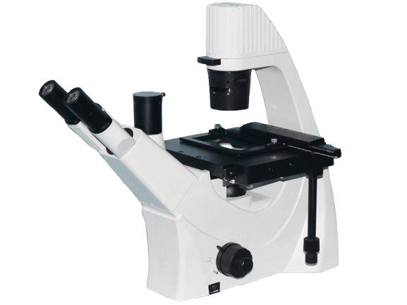
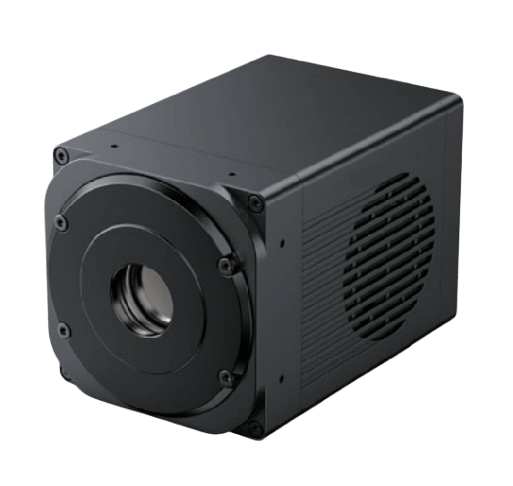
Because our cells are adherent and cultured in multi-well plates with liquid media, imaging from above is impossible. We built our entire rig around a stable inverted optical path, centered on the Venuslab IBM Inverted Microscope. This configuration allows the objective lenses to approach from the bottom of the culture dish, giving us the short working distance needed for high-resolution imaging while leaving the top accessible for environmental control.
2. Sustaining the Microenvironment
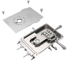
A standard microscope stage is essentially a cold slab of metal. If the temperature drops even slightly below 37°C, the cells will halt their migration, ruining the assay. To solve this, we integrated the ThermoPolar Stage directly onto the mechanical setup. This precision closed-loop system acts as a miniature incubator, maintaining optimal temperature and preventing focus drift caused by thermal expansion over the 48-hour imaging window.
3. High-Throughput Automation
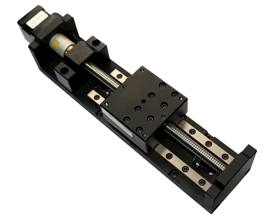
We rarely image just one well at a time. To gather statistically significant data, we need to image specific coordinates across a 96-well plate every 15 minutes. Manually moving the plate is out of the question. By retrofitting our setup with the VenusLab-Translation Axis Motorized Stage, we can program the exact X-Y coordinates of every "scratch" in the plate. The stage automatically cycles through these positions with micron-level repeatability, ensuring the exact same field of view is captured at every time point.
4. Gentle Illumination for Fluorescent Imaging
To track specific proteins during migration, our cells are tagged with green and red fluorescent proteins (GFP/RFP). Traditional mercury lamps expose cells to harsh UV radiation and excessive heat, leading to rapid photobleaching and cell death.
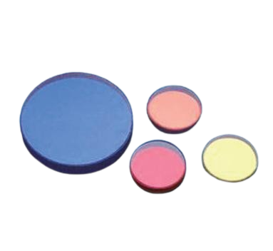
We completely replaced our arc lamps with solid-state light sources, specifically the Fiber-Coupled LED system. These are triggered electronically, meaning the sample is illuminated only for the exact millisecond the camera is exposing. To ensure clean signal separation between the green and red channels, we paired the light source with high-transmission Narrowband Interference Filters, which effectively block unwanted background noise and maximize the signal-to-noise ratio.
5. Capturing the Faint Signals
Live cells emit incredibly weak fluorescent signals, and because we use very low light intensity to protect them, the camera needs to be exceptionally sensitive. We utilize the VenusLab Scientific sCMOS Camera to capture these events. The back-illuminated sensor, known for its high quantum efficiency (QE) and low read noise, allows us to use shorter exposure times. This not only speeds up the multi-well scanning process but also further reduces phototoxicity.
Conclusion
By integrating precise environmental controls, automated stage movements, and gentle LED illumination into a stable inverted platform, we've transformed a labor-intensive observation process into a fully automated, high-throughput workflow. This setup allows us to walk away from the bench and return two days later to perfectly focused, high-contrast time-lapse data.